Valorie Mcaloon
  • Blog
  • About
  • Contact

Shoe Lifts For Leg Length Discrepancy

7/2/2017

 
Overview

leg length discrepancy can be present from birth (congenital) or acquired (a result of an injury, infection or tumor). Some of the conditions that can cause limb problems in a child or young adult include congenital conditions present from birth. Osteogenesis imperfecta,. Bow legs. Knock knees. Neurofibromatosis. Arthritis. Infections of the bones and tumors. Injuries involving the growth center of the bone. There may also be deformities that are a result of the soft tissues and not the bones, such as with arthrogryposis and burns.Leg Length Discrepancy

Causes

Some limb-length differences are caused by actual anatomic differences from one side to the other (referred to as structural causes). The femur is longer (or shorter) or the cartilage between the femur and tibia is thicker (or thinner) on one side. There could be actual deformities in one femur or hip joint contributing to leg length differences from side to side. Even a small structural difference can amount to significant changes in the anatomy of the limb. A past history of leg fracture, developmental hip dysplasia, slipped capital femoral epiphysis (SCFE), short neck of the femur, or coxa vara can also lead to placement of the femoral head in the hip socket that is offset. The end-result can be a limb-length difference and early degenerative arthritis of the hip.

Symptoms

Often there are few or no symptoms prior to the age of 25-35. The most common symptom is chronic lower back pain, but also is frequently middle and upper back pain. Same-sided and repeated injury or pain to the hip, knee and/or ankle is also a hallmark of a long-standing untreated LLD. It is not uncommon to have buttock or radiating hip pain that is non-dermatomal (not from a disc) and tends to go away when lying down.

Diagnosis

There are several orthopedic tests that are used, but they are rudimentary and have some degree of error. Even using a tape measure with specific anatomic landmarks has its errors. Most leg length differences can be seen with a well trained eye, but I always recommend what is called a scanagram, or a x-ray bone length study (see picture above). This test will give a precise measurement in millimeters of the length difference.

Non Surgical Treatment

You and your physician should discuss whether treatment is necessary. For minor LLDs in adults with no deformity, treatment may not be necessary. Because the risks may outweigh the benefits, surgical treatment to equalize leg lengths is usually not recommended if the difference is less than one inch. For these small differences, your physician may recommend a shoe lift. A lift fitted to the shoe can often improve your walking and running, as well as relieve back pain caused by LLD. Shoe lifts are inexpensive and can be removed if they are not effective. They do, however, add weight and stiffness to the shoe.

LLD Shoe Inserts

shoe lifts for men's shoes

Surgical Treatment

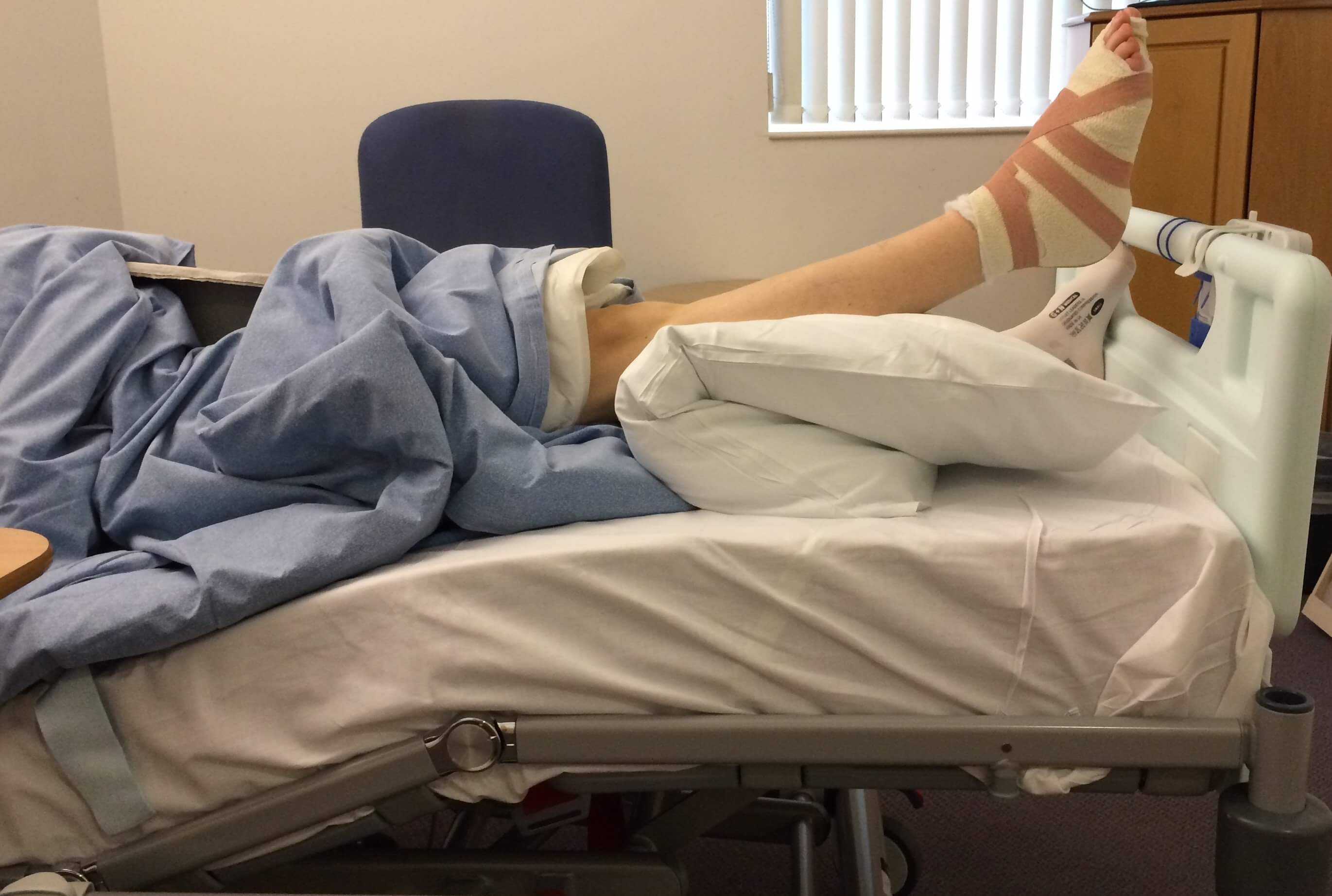
The bone is lengthened by surgically applying an external fixation device to the leg. The external fixator, a scaffold-like frame, is connected to the bone with wires, pins, or both. A small crack is made in the bone and the frame creates tension when the patient or family member turns its dial. This is done several times each day. The lengthening process begins approximately five to 10 days after surgery. The bone may lengthen 1 millimeter per day, or approximately 1 inch per month. Lengthening may be slower in a bone that was previously injured. It may also be slower if the leg was operated on before. Bones in patients with potential blood vessel abnormalities, such as cigarette smokers, may also need to be lengthened more slowly. The external fixator is worn until the bone is strong enough to support the patient safely. This usually takes about three months for each inch. Factors such as age, health, smoking and participation in rehabilitation can affect the amount of time needed.

Fallen Arches What To Look For

7/1/2017

 
Overview

Flat Foot

Fallen arches, or flatfoot, is a condition in which the arch on the inside of the foot is flat and the entire sole of the foot rests on the ground. It affects about 40% of the general population. Although flat feet in themselves are not usually problematic, they can create problems in the feet, hips, ankles and knees. Pain may be experienced in the lower back if there are alignment problems and if the individual is engaged in a lot of heavy, high impact activities that put stress on the bones and muscles in the lower legs. The arches of most individuals are fully developed by the age of 12 to 13. While some people are born with flat arches, for others the arches fall over time. The tibial tendon, which runs along the inside of the ankle from above the ankle to the arch, can weaken with age and with heavy activity. The posterior tendon, main support structure for the arch, can become inflamed (tendonitis) or even tear if overloaded. For women, wearing high heels can affect the Achilles tendon and alter the structure and function of the ankle. The posterior tibial tendon may compensate for this stress and break down, causing the arches to fall. Obesity is another contributing factor, as well as a serious injury to the ankle or foot, arthritis and bad circulation such as occurs with diabetes.

Causes

Flat feet can be caused by injury, aging, and weight gain. They can cause pain in the feet and may lead to pain in other parts of the body such as the ankles, knees, or hips. For this reason, it behooves us to treat fallen arches. The question becomes how to do so.

Symptoms

Having flat feet can be painless and is actually normal in some people. But others with flat feet experience pain in the heel or arch area, difficulty standing on tiptoe, or have swelling along the inside of the ankle. They may also experience pain after standing for long periods of time or playing sports. Some back problems can also be attributed to flat feet.

Diagnosis

An examination of the foot is enough for the health care provider to diagnose flat foot. However, the cause must be determined. If an arch develops when the patient stands on his or her toes, the flat foot is called flexible and no treatment or further work-up is necessary. If there is pain associated with the foot or if the arch does not develop with toe-standing, x-rays are necessary. If a tarsal coalition is suspected, a CT scan is often ordered. If a posterior tibial tendon injury is suspected, your health care provider may recommend an MRI.

deelsonheels

Non Surgical Treatment

If you have flat feet and foot pain, especially if one foot is flatter than the other, you should have an evaluation by an orthopedic surgeon . You may have a problem with the posterior tibial tendon , the main tendon that supports the arch. Factors that can contribute to this problem are obesity, diabetes , high blood pressure , certain types of arthritis and athletic overuse. In some cases a shoe insert/orthotic can be used to alleviate the symptoms of flat feet.

Surgical Treatment

Acquired Flat Feet

Fallen arches may occur with deformities of the foot bones. Tarsal coalition is a congenital condition in which the bones of the foot do not separate from one another during development in the womb. A child with tarsal coalition exhibits a rigid flat foot, which can be painful, notes the patient information website eOrthopod. Surgery may prove necessary to separate the bones. Other foot and ankle conditions that cause fallen arches may also require surgery if noninvasive treatments fail to alleviate pain and restore normal function.

After Care

Patients may go home the day of surgery or they may require an overnight hospital stay. The leg will be placed in a splint or cast and should be kept elevated for the first two weeks. At that point, sutures are removed. A new cast or a removable boot is then placed. It is important that patients do not put any weight on the corrected foot for six to eight weeks following the operation. Patients may begin bearing weight at eight weeks and usually progress to full weightbearing by 10 to 12 weeks. For some patients, weightbearing requires additional time. After 12 weeks, patients commonly can transition to wearing a shoe. Inserts and ankle braces are often used. Physical therapy may be recommended. There are complications that relate to surgery in general. These include the risks associated with anesthesia, infection, damage to nerves and blood vessels, and bleeding or blood clots. Complications following flatfoot surgery may include wound breakdown or nonunion (incomplete healing of the bones). These complications often can be prevented with proper wound care and rehabilitation. Occasionally, patients may notice some discomfort due to prominent hardware. Removal of hardware can be done at a later time if this is an issue. The overall complication rates for flatfoot surgery are low.

What Are The Causes Of Heel Serious Pain

6/30/2017

 
Overview

Painful Heel

The plantar fascia is a broad fan shaped strap of strong body tissue which stretches from the bottom of the heel bone to the ball of the foot. It helps to hold the foot bones and joints in place. When it is over stressed (over stretched) typical symptoms occur. The heels hurt most of all first thing in the morning or after a period of rest. The heels are also very sore after standing for a long time.

Causes

Heel pain can have many causes but the vast majority is caused by plantar fasciitis. Plantar means, ?bottom of the foot.? Fascia is a ligament or ?bundle? of ligaments. The plantar fascia is the thick ligament that helps to hold up the foot and provide spring in our step. Plantar fasciitis is an inflammation of the plantar fascia and causes more than 90% of heel pain among adults in the US. Plantar fasciitis can be acute, that is, as simple strain of the ligament but often is chronic, hanging on for months if not years. Why does that happen? The answer is poor foot mechanics, the foot sinking down too far allowing the plantar fascia to overstretch with each step taken.

Symptoms

The heel can be painful in many different ways, depending on the cause. Plantar fasciitis commonly causes intense heel pain along the bottom of the foot during the first few steps after getting out of bed in the morning. This heel pain often goes away once you start to walk around, but it may return in the late afternoon or evening. Although X-ray evidence suggests that about 10% of the general population has heels spurs, many of these people do not have any symptoms. In others, heel spurs cause pain and tenderness on the undersurface of the heel that worsen over several months. In a child, this condition causes pain and tenderness at the lower back portion of the heel. The affected heel is often sore to the touch but not obviously swollen. Bursitis involving the heel causes pain in the middle of the undersurface of the heel that worsens with prolonged standing and pain at the back of the heel that worsens if you bend your foot up or down. Pump bump, this condition causes a painful enlargement at the back of the heel, especially when wearing shoes that press against the back of the heel. Heel bruises, like bruises elsewhere in the body, may cause pain, mild swelling, soreness and a black-and-blue discoloration of the skin. Achilles tendonitis, this condition causes pain at the back of the heel where the Achilles tendon attaches to the heel. The pain typically becomes worse if you exercise or play sports, and it often is followed by soreness, stiffness and mild swelling. A trapped nerve can cause pain, numbness or tingling almost anywhere at the back, inside or undersurface of the heel. In addition, there are often other symptoms, such as swelling or discoloration - if the trapped nerve was caused by a sprain, fracture or other injury.

Diagnosis

The diagnosis of heel pain and heel spurs is made by a through history of the course of the condition and by physical exam. Weight bearing x-rays are useful in determining if a heel spur is present and to rule out rare causes of heel pain such as a stress fracture of the heel bone, the presence of bone tumors or evidence of soft tissue damage caused by certain connective tissue disorders.

Non Surgical Treatment

Treatment for plantar fasciitis - the vast majority of patients recover with conservative treatments (designed to avoid radical medical therapeutic measures or operative procedures) within months. Heel with ice-pack. Home care such as rest, ice-pack use, proper-fitting footwear and foot supports are often enough to ease heel pain. Non-steroidal anti-inflammatory drugs (NSAIDs) - medications with analgesic (pain reducing), antipyretic (fever reducing) effects. In higher doses they also have anti-inflammatory effects, they reduce inflammation (swelling). Non-steroidal distinguishes NSAIDs from other drugs which contain steroids, which are also anti-inflammatory. NSAIDs are non-narcotic (they do not induce stupor). For patients with plantar fasciitis they may help with pain and inflammation. Corticosteroids, a corticosteroid solution is applied over the affected area on the skin; an electric current is used to help absorption. Alternatively, the doctor may decide to inject the medication. However, multiple injections may result in a weakened plantar fascia, significantly increasing the risk of rupture and shrinkage of the fat pad covering the heel bone. Some doctors may use ultrasound to help them make sure they have injected in the right place Corticosteroids are usually recommended when NSAIDs have not helped. Physical therapy, a qualified/specialized physical therapist (UK: physiotherapist) can teach the patient exercises which stretch the plantar fascia and Achilles tendon, as well as strengthening the lower leg muscles, resulting in better stabilization of the ankle and heel. The patient may also be taught how to apply athletic taping, which gives the bottom of the foot better support. Night splints, the splint is fitted to the calf and foot; the patient keeps it on during sleep. Overnight the plantar fascia and Achilles tendon are held in a lengthened position; this stretches them. Orthotics, insoles and orthotics (assistive devices) can be useful to correct foot faults, as well as cushioning and cradling the arch during the healing process. Extracorporeal shock wave therapy, sound waves are aimed at the affected area to encourage and stimulate healing. This type of therapy is only recommended for chronic (long-term) cases, which have not responded to conservative therapy.

Surgical Treatment

Only a relatively few cases of heel pain require surgery. If required, surgery is usually for the removal of a spur, but also may involve release of the plantar fascia, removal of a bursa, or a removal of a neuroma or other soft-tissue growth.

how to get rid of heel spurs

Prevention

Pain On The Heel

Preventing heel pain is crucial to avoid pain that can easily interrupt a busy or active lifestyle. Athletes can prevent damage by stretching the foot and calf both before and after an exercise routine. The plantar fascia ligament can be stretched by using a tennis ball or water bottle and rolling it across the bottom of the foot. With regular stretching, the stretching and flexibility of tissue through the foot can be significantly improved, helping to prevent damage and injury. Athletes should also ease into new or more difficult routines, allowing the plantar fascia and other tissue to become accustomed to the added stress and difficulty. Running up hills is also common among athletes in their routines. However, this activity should be reduced since it places an increased amount of stress on the plantar fascia and increases the risk of plantar fasciitis. Maintaining a healthy weight is also an essential heel pain prevention technique. Obesity brings additional weight and stress on the heel of the foot, causing damage and pain in the heel as well as in other areas of the foot.

Mortons Neuroma Solutions

6/5/2017

 
Overview

Morton neuromaMorton's neuroma is an inflammation of the nerves in the foot that go to the toes. Although the name includes the word ?neuroma,? it is not really a tumor. It can affect any of the toes in the foot. However, it most often affects the nerves that run between the third and fourth, or second and third toes.
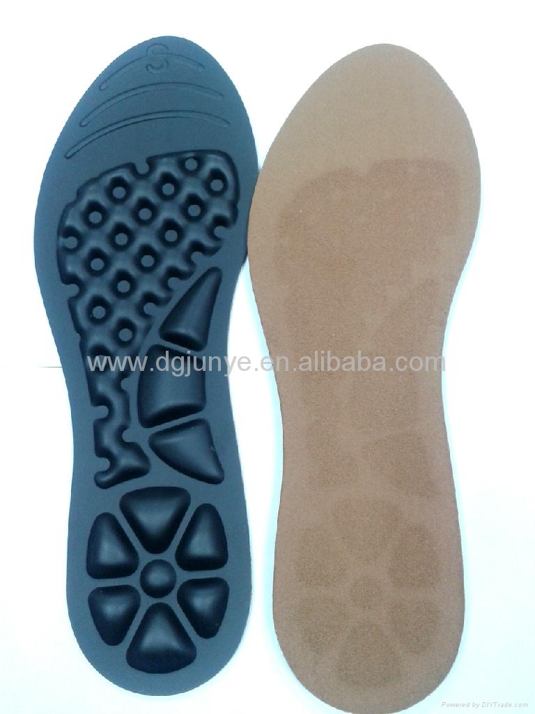
Causes

Morton's Neuroma is a foot condition caused from an abnormal function of the foot that leads to bones squeezing a nerve usually between the 3rd and 4th metatarsal heads. Symptoms of Morton's Neuroma often occur during or after you have been placing significant pressure on the forefoot area, while walking, standing, jumping, or sprinting. This condition can also be caused by footwear selection. Footwear with pointed toes and/or high heels can often lead to a neuroma. Constricting shoes can pinch the nerve between the toes, causing discomfort and extreme pain.

Symptoms

If you have a Morton's neuroma, you will probably have one or more of these symptoms. Tingling, burning, or numbness. A feeling that something is inside the ball of the foot, or your sock is bunched up. Pain that is relieved by removing your shoes. A Morton's Neuroma often develops gradually. At first the symptoms may occur only occasionally, when wearing narrower shoes or performing certain activities. The symptoms may go away temporarily by massaging the foot or by avoiding aggravating shoes or activities. Over time the symptoms progressively worsen and may persist for several days or weeks. The symptoms become more intense as the neuroma enlarges and the temporary changes in the nerve become permanent.

Diagnosis

A GP (general practitioner, primary care physician) or a podiatrist (foot specialist doctor) will ask the patient to describe the pain as well as its intensity, when symptoms started, what types of shoes are worn, as well as some questions about their job, lifestyle and hobbies. The doctor will then examine the foot and try to locate the affected nerve. This may involve attempting to reproduce symptoms by manipulating the foot. In order to get a detailed image of the inside of the food, one of the following scans may be ordered. X-ray, this is a type of high-energy radiation. In low doses they are used to diagnose diseases and condition by making pictures of the inside of the body. In higher doses they are used to treat cancer. This procedure is non-invasive and painless. Ultrasound scan, high frequency sound waves are pointed at a specific part of the body, which in this case is the foot. The ultrasound waves bounce of tissues; their echoes are then converted into a picture, called a sonogram. This allows the doctor to get an inside view of the foot. This procedure is non-invasive and painless. MRI (magnetic resonance imagining) a device that uses a magnetic field and radio waves to create detailed images of the body. Unlike CT scanning or general x-ray studies, no ionizing radiation is involved with an MRI. This procedure is non-invasive and painless. The doctor will have to rule out other conditions which may have similar symptoms, including capsulitis, bursitis, or Freiberg's disease.

Non Surgical Treatment

Properly resting the foot in addition to the use of appropriate footwear including, as necessary, pads and arch supports, often brings relief from Morton?s Neuroma, without resorting to surgery. A physician may also recommend anti-inflammatory drugs, cortisone injections and, should the condition fail to resolve itself, surgery.Morton

Surgical Treatment

Surgery. This is the last and most permanent course of action. This surgery is used as a last resort as it often comes with a series of side affects including the risk of making the pain worse. This surgery can be performed by Orthopedic surgeons as well as Podiatric surgeons.

Prevention

While Morton?s Neuroma has been an ongoing topic of clinical investigation, the condition is in some cases difficult to either treat or prevent. Experimental efforts involving the injection of muscle or bone with chemicals such as alcohol, as well as suturing, and covering affected areas with silicone caps have been attempted, with varying success.

Are Shoe Lifts The Best Solution To Leg Length Imbalances

2/20/2016

 
There are actually not one but two different types of leg length discrepancies, congenital and acquired. Congenital indicates you are born with it. One leg is structurally shorter compared to the other. Through developmental phases of aging, the human brain senses the stride pattern and recognizes some variance. The human body usually adapts by tilting one shoulder over to the "short" side. A difference of less than a quarter inch is not really irregular, require Shoe Lifts to compensate and mostly won't have a serious effect over a lifetime.

Leg Length Discrepancy Shoe Lifts

Leg length inequality goes mainly undiscovered on a daily basis, yet this condition is very easily corrected, and can eliminate quite a few instances of lower back pain.

Treatment for leg length inequality typically involves Shoe Lifts. These are generally very reasonably priced, regularly priced at under twenty dollars, in comparison to a custom orthotic of $200 if not more. When the amount of leg length inequality begins to exceed half an inch, a whole sole lift is generally the better choice than a heel lift. This prevents the foot from being unnecessarily stressed in an abnormal position.

Back pain is easily the most widespread ailment affecting men and women today. Over 80 million men and women suffer from back pain at some stage in their life. It's a problem which costs companies millions yearly on account of lost time and productivity. New and improved treatment solutions are constantly sought after in the hope of minimizing the economical influence this condition causes.

Shoe Lift

People from all corners of the world suffer the pain of foot ache due to leg length discrepancy. In these situations Shoe Lifts are usually of beneficial. The lifts are capable of alleviating any discomfort in the feet. Shoe Lifts are recommended by numerous skilled orthopaedic practitioners".

So as to support the human body in a well-balanced manner, the feet have a crucial task to play. In spite of that, it can be the most neglected region of the body. Many people have flat-feet which means there is unequal force placed on the feet. This causes other body parts including knees, ankles and backs to be affected too. Shoe Lifts ensure that the right posture and balance are restored.

What May Cause Posterior Calcaneal Spur

9/29/2015

 
Calcaneal Spur

Overview

A heel spur is a buildup of calcium or a bone hook on the heel bone. This is typically the source of most heel pain. It usually takes an X-ray to see the heel spur protruding from the heel. Without proper heel spur treatment, a heel spur cause inflammation and lead to other ailments like plantar fasciitis and Achilles tendonitis. It is important to be examined by an orthopedic specialist.

Causes

This condition is a constellation of many causes; overweight, ill fitting shoes, bio-mechanical problems (mal-alignment of the heel), gout, pronation (a complex motion including outward rotation of the heel and inward rotation of the ankle) and rheumatoid arthritis are some of the causes of heel pain.

Calcaneal Spur

Symptoms

The spur itself is not painful, however, if it is sharp and pointed it can poke into soft tissue surrounding the spur itself. As the bone spur irritates the tissue, inflammation and bruising can occur leading to heel pain. Heel spurs can affect your ability to do your usual work and/or activities, and can also trap and irritate the nerves in your heel area. They can change the way you walk, and can lead to knee, hip and low back injuries. If severe, they may require medical intervention.

Diagnosis

Diagnosis of a heel spur can be done with an x-ray, which will be able to reveal the bony spur. Normally, it occurs where the plantar fascia connects to the heel bone. When the plantar fascia ligament is pulled excessively it begins to pull away from the heel bone. When this excessive pulling occurs, it causes the body to respond by depositing calcium in the injured area, resulting in the formation of the bone spur. The Plantar fascia ligament is a fibrous band of connective tissue running between the heel bone and the ball of the foot. This structure maintains the arch of the foot and distributes weight along the foot as we walk. However, due to the stress that this ligament must endure, it can easily become damaged which commonly occurs along with heel spurs.

Non Surgical Treatment

FIRST, Reduce the acute pain. This is done by a combination of several things; injection of a synthetic relative of cortisone into the heel, a prescription of anti-inflammatory pills to reduce inflammation, physical therapy and a special heel pad. About 50% of the time, these treatments will permanently relieve the pain. In the other 50%, the pain becomes recurrent, and the treatment proceeds to Stage II. SECOND, Recurrent, painful heel spur is caused by the tug and pull of the plantar fascia ligament on the heel bone with each step. When the pain is recurrent, arch supports are made to prevent sagging of the arch. The arch supports are custom-made according to the size and shape of the feet. This prevents the arch from sagging and the ligament from tugging and pulling on the heel bone. The inflammation and pain eventually go away as the first phase of treatment is continued along with the arch supports, although the spur itself remains. THIRD, Surgery to remove the spur is possible and is usually done as Day Surgery.

Surgical Treatment

In a small number of cases (usually less than 5 percent), patients may not experience relief after trying the recommendations listed above. It is important that conservative treatments (such as those listed above) be performed for AT LEAST a year before considering surgery. Time is important in curing the pain from heel spurs, and insufficient treatment before surgery may subject you to potential complications from the procedure. If these treatments fail, your doctor may consider an operation to loosen the plantar fascia, called a plantar fascia release.

Prevention

To prevent this condition, wearing shoes with proper arches and support is very important. Proper stretching is always a necessity, especially when there is an increase in activities or a change in running technique. It is not recommended to attempt working through the pain, as this can change a mild case of heel spurs and plantar fascitis into a long lasting and painful episode of this condition.

Tips To Treat Calcaneal Spur

9/27/2015

 
Heel Spur

Overview

A heel spur is a hooked bony growth protruding from the calcaneus or heel bone. It often occurs alongside plantar fasciitis, and as such the two conditions are often confused, however they are not the same.

Causes

The plantar fascia is a big strong ligament on the bottom of the foot, starting at the bottom of the heel bone and running into the ball of the foot. As the arch of the foot becomes weak, it sags slightly with each step and this causes the plantar fascia to tug and pull at the heel bone with each step. Over a period of time, a spur forms where this big strong ligament tugs and pulls at the heel bone. Soon, inflammation (swelling) starts around this spur and the pain becomes almost unbearable. (Sometimes heel spurs may be present without being painful if no inflammation is present).

Inferior Calcaneal Spur

Symptoms

With heel spurs, people often talk about a dull ache which is felt most of the time with episodes of a sharp pain in the center of the heel or on the inside margin of the heel. Often the pain is worse on first rising in the morning and after rest and is aggravated by prolonged weight bearing and thin-soled shoes.

Diagnosis

Most patients who are suffering with heel spurs can see them with an X-ray scan. They are normally hooked and extend into the heel. Some people who have heel spur may not even have noticeable symptoms, although could still be able to see a spur in an X-ray scan.

Non Surgical Treatment

Get some rest. You need to stay off of your aching foot as much as possible for at least a week. Think about possible causes of the problem while you're resting and figure out how you can make some changes. Some actions that can contribute to heel spurs include running too often or running on hard surfaces such as concrete, tight calf muscles, shoes with poor shock absorption. Ease back into your activities. In many cases, you'll be in too much pain to go ahead with a strenuous exercise routine that puts pressure or impact on your heel. Listen to your body and switch to different activities such as swimming or riding a bike until your heel spurs improve.

Surgical Treatment

Have surgery if no other treatments work. Before performing surgery, doctors usually give home treatments and improved footwear about a year to work. When nothing else eases the pain, here's what you need to know about surgical options. Instep plantar fasciotomy. Doctors remove part of the plantar fascia to ease pressure on the nerves in your foot. Endoscopy. This surgery performs the same function as an instep plantar fasciotomy but uses smaller incisions so that you'll heal faster. However, endoscopy has a higher rate of nerve damage, so consider this before you opt for this option. Be prepared to wear a below-the-knee walking cast to ease the pain of surgery and to speed the healing process. These casts, or "boots," usually work better than crutches to speed up your recovery time.

Prevention

To prevent this condition, wearing properly fitted shoes with good arch support is very important. If a person is overweight, weight loss can help diminish stress on the feet and help prevent foot problems. For those who exercise frequently and intensely, proper stretching is always necessary, especially when there is an increase in activities or a change in running technique. It is not recommended to attempt to work through the pain, as this can change a mild case of heel spurs and plantar fascitis into a long-lasting and painful episode of the condition.

Bursitis Top Of Feet Therapy

8/26/2015

 
Overview

A bursa is a fluid-filled sac. There are hundreds of deep and superficial bursae throughout the human body. They are typically located near major joints. Bursae are situated between bone and soft tissues like tendons, ligaments, muscles, and skin. They serve as a cushioning pad to absorb shock. The fluid within the sac is secreted to assist with friction-free movement as the soft tissues move across a bony area.

Causes

A bursa is a fluid-filled sac that acts as a cushion and a lubricant between tendons and muscles sliding over bone. There are bursas around most large joints in the body, including the ankle. The retrocalcaneal bursa is located in the back of the ankle by the heel, where the large Achilles tendon connects the calf muscles to the heel bone (calcaneus). Repetitive or over use of the ankle, for instance by doing excessive walking, running or jumping, can cause this bursa to become irritated and inflamed. This condition is commonly associated with Achilles tendonitis. Sometimes retrocalcaneal bursitis may be mistaken for achilles tendonitis. Those at risk for this condition include people just starting aggressive exercise regimens or having some other sudden increase in activity without proper conditioning.

Symptoms

Medical experts strongly recommend that you consult a doctor if you have any of the symptoms below. Disabling joint pain that prevents you from doing your daily activities. Pain that lasts for more than two weeks. Excessive swelling, redness, bruising or a rash around the painful joint. Sharp or shooting pain, especially when you exercise or do something more strenuous. A fever. Any of the above could be a sign of infection, a condition such as arthritis or a more serious injury such as a tendon tear that may require medical attention.

Diagnosis

Medical examination is not necessarily required in light cases where the tenderness is minimal. In all cases where smooth improvement is not experienced, medical attention should be sought as soon as possible to exclude a (partial) rupture of the Achilles tendon or rupture of the soleus muscle. This situation is best determined by use of ultrasound scanning, as a number of injuries requiring treatment can easily be overlooked during a clinical examination (Ultrasonic image). Ultrasound scanning enables an evaluation of the extent of the change in the tendon, inflammation of the tendon (tendinitis), development of cicatricial tissue (tendinosis), calcification, inflammation of the tissue surrounding the tendon (peritendinitis), inflammation of the bursa (bursitis), as well as (partial) rupture.

Non Surgical Treatment

In addition to R.I.C.E., there are a number of other treatments to reduce swelling and any associated pain or discomfort due to heel bursitis. Orthotics or change of footwear. Wearing an orthotic device such as a heel insert can encourage better mechanics in the foot and reduce irritation of the retrocalcaneal bursa. Some people do not need special orthotics but simply need to stop wearing shoes with rigid heel and ankle construction and instead wear more supportive, comfortable shoes. Shoes with an "Achilles notch," a groove in the collar at the back of the shoe to protect the Achilles tendon, can be particularly helpful. (Almost all running shoes are designed with an Achilles notch.) Stretching and physical therapy. Stretching the Achilles tendon often helps alleviate pain. Once the pain is resolved it is important for the patient to continue a regular stretching program. Regular stretching reduces the chance of recurrence.

Surgical Treatment

Bursectomy is a surgical procedure used to remove an inflamed or infected bursa, which is a fluid-filled sac that reduces friction between tissues of the body. Because retrocalcaneal bursitis can cause chronic inflammation, pain and discomfort, bursectomy may be used as a treatment for the condition when it is persistent and cannot be relived with other treatments. During this procedure, a surgeon makes small incisions so that a camera may be inserted into the joint. This camera is called an arthroscope. Another small incision is made so that surgical instruments can be inserted to remove the inflamed bursa.

Prevention

You can help to prevent heel pain and bursitis by maintaining a healthy weight, by warming up before participating in sports and by wearing shoes that support the arch of the foot and cushion the heel. If you are prone to plantar fasciitis, exercises that stretch the Achilles tendon (heel cord) and plantar fascia may help to prevent the area from being injured again. You also can massage the soles of your feet with ice after stressful athletic activities. Sometimes, the only interventions needed are a brief period of rest and new walking or running shoes.

Contracted Toes

6/30/2015

 
HammertoeOverview

Hammer toe is defined as a deformity in the toe where part of the toe is permanently bent downward resembling a hammer. Two related conditions are mallet toe and claw toe which effect different toe joints in slightly different ways. The key difference is that hammertoes tends to effect the middle joint in the toe (note: not the middle toe, the middle toe joint). The Hammer toes disease is usually associated with the second largest toe but can effect the third or fourth toe as well. Mallet toe effects the uppermost toe joint whereas claw toe is caused by the tow being held in a cramped ?claw-like? position.

Causes

Hammer toe usually affects the second toe. However, it may also affect the other toes. The toe moves into a claw-like position. The most common cause of hammer toe is wearing short, narrow shoes that are too tight. The toe is forced into a bent position. Muscles and tendons in the toe tighten and become shorter. Hammer toe is more likely to occur in women who wear shoes that do not fit well or have high heels and children who keep wearing shoes they have outgrown. The condition may be present at birth (congenital) or develop over time. In rare cases, all of the toes are affected. This may be caused by a problem with the nerves or spinal cord.

Hammer ToeSymptoms

Here is a look at some of the symptoms hammertoe can cause. They include hammer-like or claw-like appearance of the toe. Pain when walking or moving the foot. Difficulty moving the toe. Corns may form on top of the toe. Callus may form on the sole of the foot. During the initial stages, you may be able to manually straighten your toe. This is called a flexible hammertoe. But as time passes, the toe will not move as easily and will continue to look like a hammer. Pressure and irritation over the joint can cause a blister to develop and become a corn over time. These corns have the potential to become infected and cause additional symptoms such as redness, bleeding, and difficulty wearing shoes and socks. Corns are the main cause of pain when hammertoes are developing.

Diagnosis

First push up on the bottom of the metatarsal head associated with the affected toe and see if the toe straightens out. If it does, then an orthotic could correct the problem, usually with a metatarsal pad. If the toe does not straighten out when the metatarsal head is pushed up, then that indicates that contracture in the capsule and ligaments (capsule contracts because the joint was in the wrong position for too long) of the MTP joint has set in and surgery is required. Orthotics are generally required post-surgically.

Non Surgical Treatment

Hammer toes may be effectively corrected in different ways. Treatments can be non-invasive and involve physical therapy along with the advice that the person not wear any more shoes that restrict appropriate space for their toes. Appropriate shoes for people who want to avoid hammer toes, or for people who already have them, should be at least half an inch longer than the person's longest toe. High-heeled shoes are something to definitely avoid.

Surgical Treatment

he basis for hammer toe surgery most often involves removing s portion of bone within the toe, to reduce the joint contracture. Depending on the direction the toe is deviated, soft tissue procedures may be necessary along with pinning the toe with a surgical wire.

Hammer Toe Treatment In Your Own Home

6/27/2015

 
HammertoeOverview

Generally a hammertoe or mallet toe is caused by wearing high heels or shoes that are too small around the toe area, so it?s no surprise that it is mostly women who suffer from them. A hammertoe has a bend in the middle joint of the toe whereas a mallet toe has a bend in the upper joint of the affected toe. The way someone walks (gait) can also lead to the formation of hammertoes and mallet toes as can overuse and injury. Sometimes a deep blister will form over the bent joint and often after some time calluses and corns will develop on the affected toe joint. People with arthritis, diabetes or neuromuscular conditions are also more likely to develop a hammer toe or mallet toe.

Causes

The constant pressure a woman's foot receives in high-heeled shoes due to the force of gravity causes their feet to naturally slide down and press on the lowest point of the shoe so they are not able to receive enough space and stretch out. The result is an eventual distortion of the woman's toes. The deformity comes as a result of the shortening of muscles inside the toes because the toes become used to being in a Hammer toes bent position, prompting the muscles to fail to extend any further and become tightened and curbed. At first, toes may still be stretched out if poor footwear is not being worn, yet if the habit is persistent...the person's toes will eventually become used to the position they are constantly in and muscle fibers inside them will harden and refuse to stretch.

HammertoeSymptoms

The most obvious sign of hammertoes are bent toes, other symptoms may include pain and stiffness during movement of the toe. Painful corns on the tops of the toe or toes from rubbing against the top of the shoe's toe box. Painful calluses on the bottoms of the toe or toes. Pain on the bottom of the ball of the foot. Redness and swelling at the joints.

Diagnosis

The treatment options vary with the type and severity of each hammer toe, although identifying the deformity early in its development is important to avoid surgery. Your podiatric physician will examine and X-ray the affected area and recommend a treatment plan specific to your condition.

Non Surgical Treatment

There is a variety of treatment options for hammertoe. The treatment your foot and ankle surgeon selects will depend upon the severity of your hammertoe and other factors. A number of non-surgical measures can be undertaken. Padding corns and calluses. Your foot and ankle surgeon can provide or prescribe pads designed to shield corns from irritation. If you want to try over-the-counter pads, avoid the medicated types. Medicated pads are generally not recommended because they may contain a small amount of acid that can be harmful. Consult your surgeon about this option. Changes in shoewear. Avoid shoes with pointed toes, shoes that are too short, or shoes with high heels, conditions that can force your toe against the front of the shoe. Instead, choose comfortable shoes with a deep, roomy toe box and heels no higher than two inches. Orthotic devices. A custom orthotic device placed in your shoe may help control the muscle/tendon imbalance. Injection therapy. Corticosteroid injections are sometimes used to ease pain and inflammation caused by hammertoe. Medications. Oral nonsteroidal anti-inflammatory drugs (NSAIDs), such as ibuprofen, may be recommended to reduce pain and inflammation. Splinting/strapping. Splints or small straps may be applied by the surgeon to realign the bent toe.

Surgical Treatment

If these non-invasive treatments don?t work, or if the joint is rigid, a doctor?s only recourse may be to perform surgery. During the surgery, the doctor makes an incision and cuts the tendon to release it or moves the tendon away from or around the joint. Sometimes part of the joint needs to be removed or the joint needs to be fused. Each surgery is different in terms of what is needed to treat the hammertoe. Normally after any foot surgery, patients use a surgical shoe for four to six weeks, but often the recovery from hammertoe surgery is more rapid than that. An unfortunate reality is that hammertoe can actually return even after surgery if a patient continues to make choices that will aggravate the situation. Though doctors usually explain pretty clearly what needs to be done to avoid this.
<<Previous
Powered by Create your own unique website with customizable templates.